 Fraunhofer Institute for Electronic Nano Systems
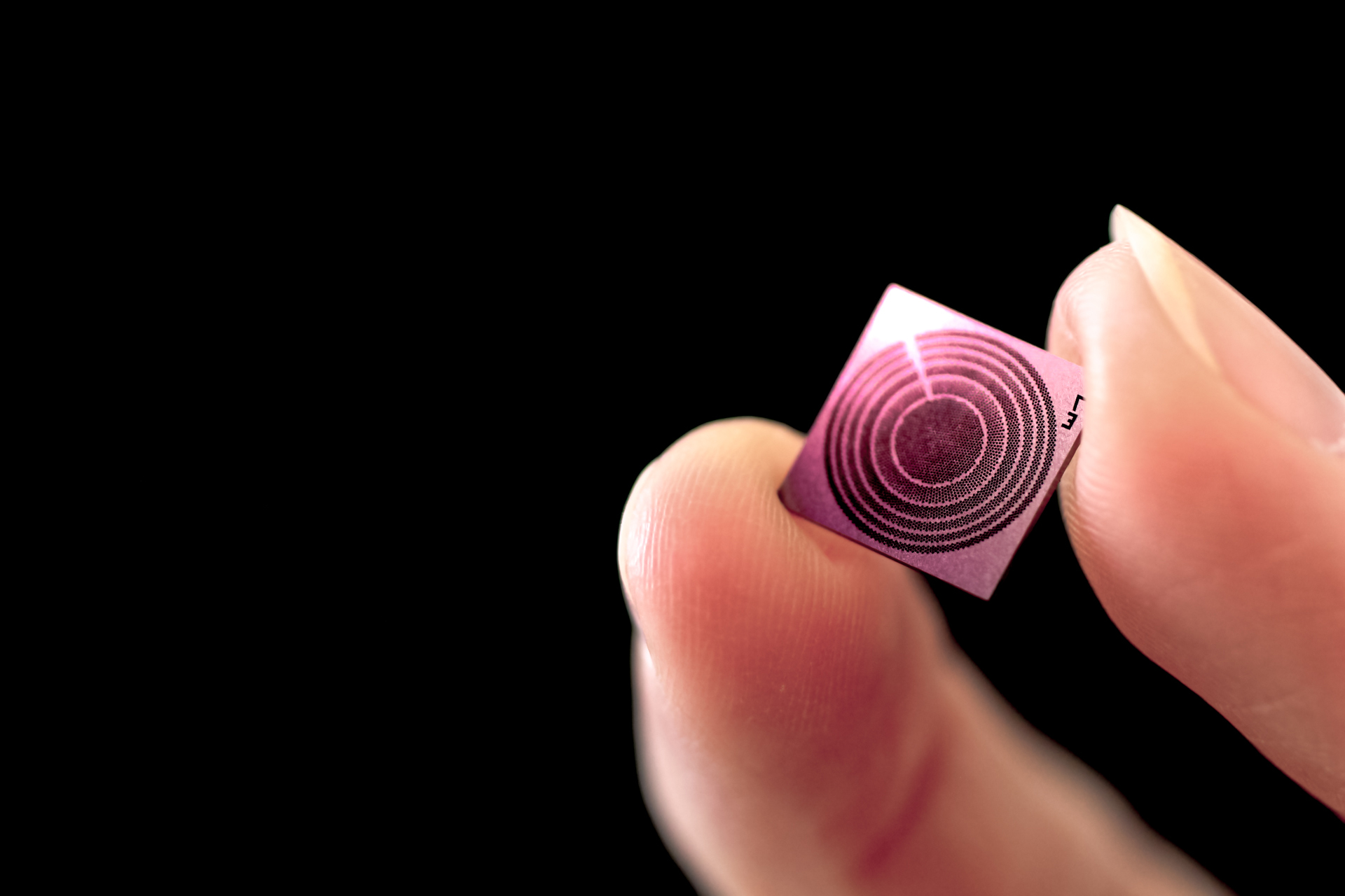
Imaging methods such as ultrasound and photoacoustics are integral components of the testing procedures used in medicine and industry today. They make it possible to visualize internal structures – from organic tissue to materials – non-invasively, without damaging or even touching them. As the key to further diagnosis, state-of-the-art methods can be used to identify even the tiniest changes and abnormalities in organs, muscles or blood vessels in real time, detect serious disease early on and initiate precision-targeted therapeutic measures. But that is not all; ultrasound methods are also used across a host of industrial applications for nondestructive testing, inspection and analysis to reveal defects and assure the quality of components. With their ultrasonic transducers, researchers from Fraunhofer ENAS are helping to unlock fascinating insights into the inside of the human body and thus improve healthcare. At the same time, they are also making it possible to trace even hidden details of technological components, enhancing safety and reliability.
Ultrasound examinations are among the most important imaging methods in the modern world today. Also known as sonography, ultrasound is used as a quick, pain-free way to gather diagnostic information pointing to serious disease or to monitor the progress of certain health conditions. Ultrasound is also helpful in industrial applications as a reliable and nondestructive way to test aspects such as material properties or detect irregularities and flaws in materials.
From echo to image: ultrasound as a window on the body in medicine
“In sonography, an ultrasonic probe is moved over the patient’s skin, in contact with the area of the body that is undergoing the scan. Electrical signals generate sound waves that penetrate deep into the body through the probe. When these waves make contact with organs or tissue, some fraction bounces back, so it is reflected like an echo. But since bones, muscles and blood vessels have different properties and structures, they vary widely in how they reflect or absorb the sound waves,” says Dr. Chris Stöckel, head of the “MEMS/NEMS Technologies” group at Fraunhofer ENAS, explaining how ultrasound works.
For example, bones are highly reflective, but ultrasonic waves pass through liquids such as blood. They do not reflect sound waves but absorb them instead. Based on these different levels of reflection and absorption, electrical impulses are generated and then used to calculate the typical black-and-white ultrasound image. Heavily reflective areas show up in white, while absorptive types of tissue are visualized in black. The 2D image of the organ or tissue scanned in this way gives doctors information about the health of that area of the body and indications of any anomalies.
Tiny technology, big impact: Micromachined ultrasonic transducers reveal delicate structures in medical diagnostics
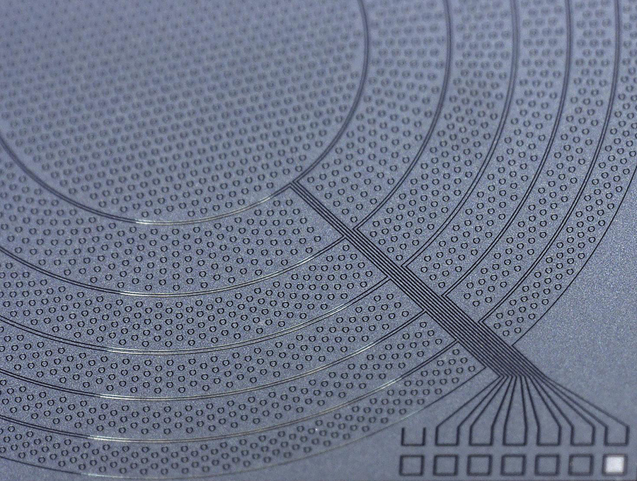
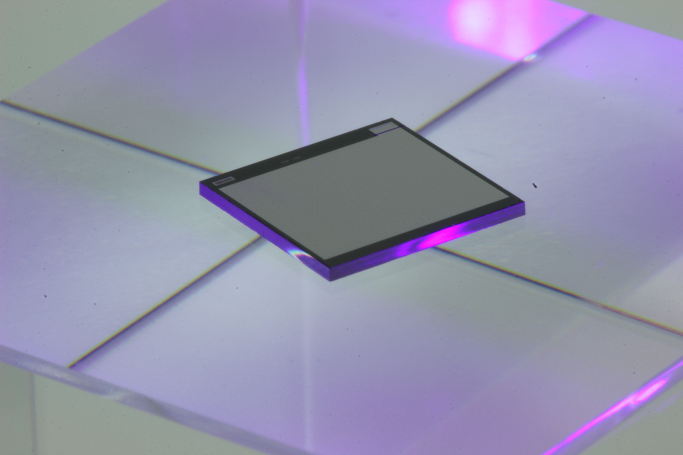
Cutting-edge high-resolution technologies are needed in order to detect even the tiniest irregularities inside the body. The researchers at Fraunhofer ENAS are working to develop one such technology. Built into ultrasonic probes, micromachined ultrasonic transducers (MUTs) from the scientists in Chemnitz allow for detection of even the finest structures.
“This is made possible by ultra-thin membranes produced on silicon wafers that are built into the inside of the ultrasonic transducer. At just one to five micrometers in thickness, they’re smaller than a red blood cell, for example. Applying an electric current starts these membranes vibrating, which creates high-frequency sound waves inaudible to the human ear. When those sound waves make contact with an object such as tissue, the signal is reflected and can be analyzed,” explains Dr. Nooshin Saeidi, head of the “Micro Acoustic Systems” group at Fraunhofer ENAS, who also working on MUT technologies.
One special feature of the ultrasonic transducers from Fraunhofer ENAS is that they can be operated capacitively or piezoelectrically. The difference lies in their structure, which typically consists of two electrodes. In capacitive micromachined ultrasonic transducers (CMUTs), an electrostatic force is created between the two electrodes. When an electric current is applied, an electric field is created, starting the membrane to vibrate and generate sound waves. In piezoelectric micromachined ultrasonic transducers (PMUTs), by contrast, piezoelectric thin films are applied directly to the membrane. These piezoelectric materials convert the electrical signals generated when current is applied into mechanical deflection, thereby causing the membrane to vibrate directly and generating ultrasonic waves.